


In the geosciences, X-rays are primarily used to examine natural minerals. The so-called powder diffraction method in the XRD laboratory of Christina Günter and Wolfgang Morgenroth has the advantage that the crystals do not need to be in their pure form; instead, a mixture of different minerals in a single sample can be detected relatively quickly and with high accuracy. “There are other, sometimes simpler methods for this,” says mineralogist Morgenroth. In certain cases, however, X-ray diffraction can serve as a reliable source for watertight evidence that stands up to scientific scrutiny.
To unlock the secrets of a tough rock like the grass-green olivine, Morgenroth and his students must first mechanically crush the rock. The mineral, found in the upper Earth’s mantle, is ground up before a small amount of fine powder can be applied to a microscope slide. The rest of the painstaking work is done almost automatically by the “Empyrean,” an XRD device from Malvern Panalytical. In the process, an X-ray beam strikes the olivine through a small window, while a detector moves steadily in a semicircle around the sample for several minutes to measure reflections of the “deflected” primary beam. This actual measurement takes place automatically, shielded behind the safety glass of the machine, which is protected from radiation to the outside. On the computer, Morgenroth can compare the measured diffraction patterns with his own data or with large databases such as those of the International Centre for Diffraction Data. The result confirms the typical parameters of the olivine unit cell.
Measuring reflections from the crystal lattice
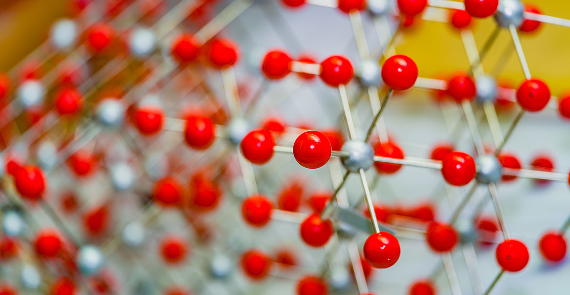
The basic principle of such measurements, postulated as early as 1912, is as old as it is ingenious. X-rays are not only suitable for scanning our bodies when we get sick or sprain an ankle. Due to their short wavelength, they are also ideal for measurements on the atomic scale. This is because in its crystalline form, as a lattice, every substance, every chemical element, scatters X-rays in space in its own unique way. “We take advantage of the fact that we irradiate a crystal that has a three-dimensionally repeating pattern,” explains Morgenroth, who trains young researchers at the X-ray diffractometer at the Institute of Geosciences. The smallest of these repeating clusters of atoms or ions is called a unit cell. Due to their periodic arrangement, all identically aligned unit cells deflect the X-ray beam at the same angle. When they interfere, the parallel waves of these reflected X-rays can amplify or cancel each other out. Thus, each crystal produces a very specific diffraction pattern in the reflection of the X-ray beam. By measuring this “fingerprint” on a detector plate or a sensor, one can deduce the composition and spatial structure of the material.
In this way, researchers can analyze sand from a limestone deposit quite precisely for its components, such as the proportions of quartz and calcium carbonate. In mixtures such as cement or building plaster, there are often significantly more components. In practical terms, such findings are particularly useful in historic preservation, for example, when the composition of old mortars that hold historic buildings together needs to be reproduced. This method is also useful for the determination of color pigments. Another area of application is the precise analysis of harmful salts that influence the weathering of a building. In the context of agricultural and environmental economics, the composition of clay minerals in soils can be determined using XRD.
Search for new chemical compounds
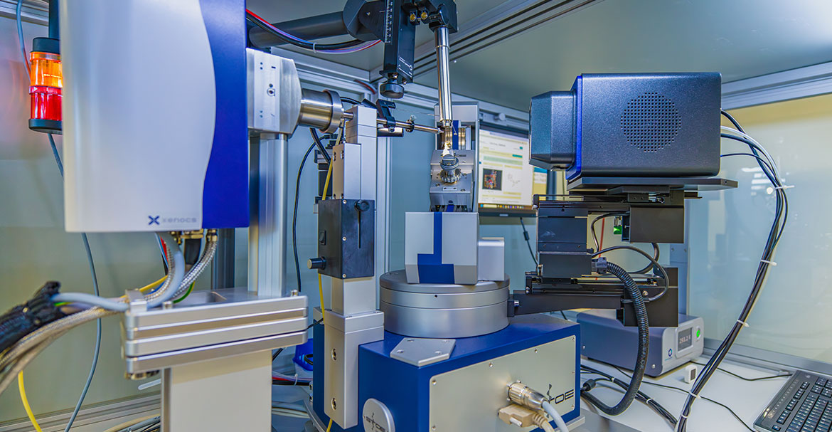
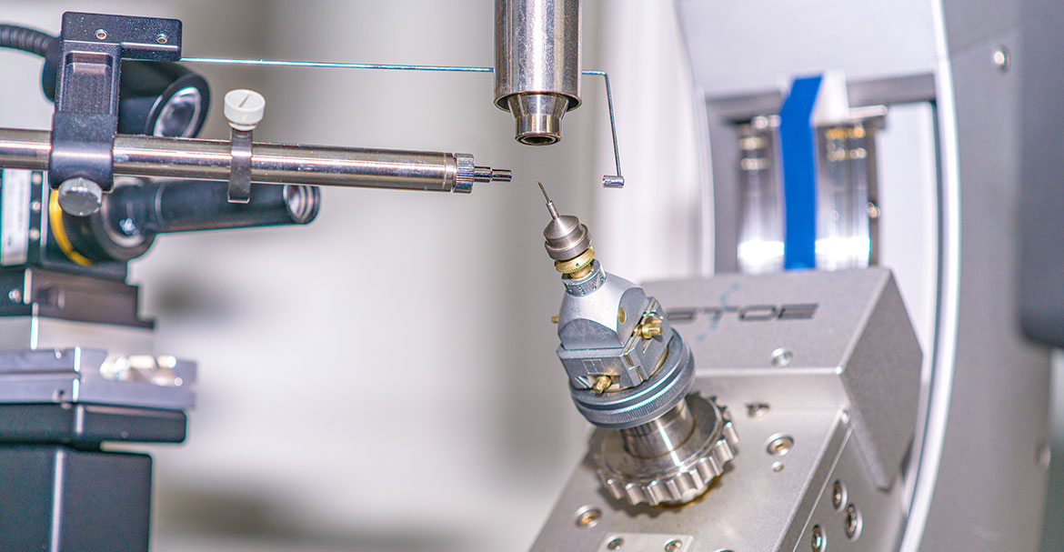
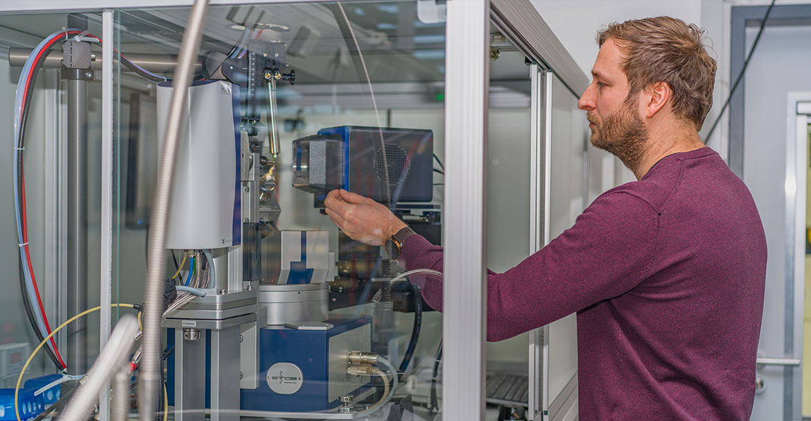
The method is of crucial importance for the research of entirely unknown compounds, particularly at the Institute of Chemistry. A low-frequency hum pervades the XRD laboratory of the institute, amidst the cooling and ventilation pipes, microscopes, and pressure vessels. It is the playground of chemist Eric Sperlich, who uses the STOE Stradivari single-crystal diffractometer here to examine tiny crystals that are no longer visible to the naked eye – often even completely novel molecules. “We’re a service center,” says the lab director. “We typically get requests from doctoral or postdoctoral students, but also from undergraduates researching the synthesis of new organic, inorganic, or hybrid materials whose molecular structure is currently unknown. The request often goes like this: ‘Tell me what this is and whether the compound I’m hoping for has actually been formed.’”
Under the microscope, Eric Sperlich sensitively secures a reddish crumb – a cobalt compound – onto a needle using special tools. A task that used to have to be done with a brush and glue or modeling clay. The sample is then centered in the XRD’s measurement area. Using a so-called four-circle goniometer, this highly complex device can irradiate this tiny speck of substance from virtually every angle. A process that takes only a few seconds and, due to its small scale, can be sensed more than observed. In the cloud, the result from 138 individual measurements is compared with the 1.4 million entries in the Cambridge Structural Database, which catalogs all chemical compounds with at least one carbon-hydrogen bond. The proverbial gist of the matter – the arrangement of atoms in the substance – is also illustrated in a computer graphic that shows exactly where each atom is located in the lattice. The great hope that always accompanies this process is the discovery of a new, previously undescribed compound whose properties bring science one step closer to solving a specific problem.
These can include components for new plastics, lithium batteries, or LEDs; materials for sensors designed to function in sodium-containing environments (such as saltwater); or even active pharmaceutical ingredients. Cristian Paz from the Universidad de La Frontera in Temuco, Chile, for example, sends extracts of medicinal plants from Chile to the institute to have them analyzed for the structure of potential active ingredients. Arranged into a lattice through chemical processes, as a so-called single crystal, organic material also reveals its internal structure. This is the prerequisite for obtaining a usable result with XRD: the substance must be in crystalline form. However, any gas or liquid can be converted into such a solid state by cooling, Sperlich says. Even if the synthesis of a new substance produces defects in the atomic lattice, the quality of such a crystal sometimes deserves the label: not beautiful, but rare. Thanks to extensive databases, computer-aided precision measurements, and powerful software, however, small defects do not distort the results too much these days; in some cases, XRD is the only way to detect them. What has been feasible in theory and practice since the early 20th century has only truly come into its own in the laboratory thanks to 21st-century computer technology and computing power. Whereas not so long ago, measuring and analyzing an unknown crystal under optimal conditions took often more than half a week, researchers today can, with a bit of luck, get their results on the very same day. Much to Sperlich’s delight, “I’m a huge fan of this method.”
Further information on X-ray crystallography at the Institute of Chemistry: https://www.uni-potsdam.de/de/kristallographie
Further information on the XRD laboratory for X-ray powder diffraction led by Christina Günter and Wolfgang Morgenroth at the Institute of Geosciences: https://www.uni-potsdam.de/de/geo/forschung/mineralogie/ausstattung/xrd-labor
This article appeared in the university magazine Portal - Eins 2026 „Inklusion“.